samedi 15 mars 2014

Burst fracture

burst fracture is a type of compression fracture which results in distruption of the posterior vertebral body cortex with retropulsion into the spinal canal. When in the thoraco lumbar level it tends to occur between T9 and L5 levels 3.  Burst fractures may be stable or unstable.

Pathology

Mechanism
It is a result of a compressive high energy injury (axial loading), much like the Jefferson fracture.  Typically it occurs following a fall, landing on the feet, from a significant height.
The intervertebral disc is driven into the vertebral body below.
All patients require a CT to assess the injury and evaluate the extent of retropulsed fragments which may enter the spinal canal.

Radiographic features

General features include
  • 'burst' vertebral body on axial CT.
  • loss of posterior vertebral height on lateral views.
  • retropulsed fragments in the spinal canal.
  • interpedicular widening

vendredi 14 mars 2014


Cardiogenic and Non-cardiogenic
Pulmonary Edema
General Considerations
  • Increase in the fluid in the lung
  • Generally, divided into cardiogenic and non-cardiogenic categories.
  • Congestive heart failure is the leading diagnosis in hospitalized patients older than 65
Pathophysiology
  • Fluid first accumulates in and around the capillaries in the interlobular septa (typically at a wedge pressure of about 15 mm Hg)
  • Further accumulation occurs in the interstitial tissues of the lungs
  • Finally, with increasing fluid, the alveoli fill with edema fluid (typically wedge pressure is 25 mm Hg or more)
Causes
  • Cardiogenic pulmonary edema.
  • Heart failure
  • Coronary artery disease with left ventricular failure.
  • Cardiac arrhythmias
  • Fluid overload -- for example, kidney failure.
  • Cardiomyopathy
  • Obstructing valvular lesions -- for example, mitral stenosis 
  • Myocarditis and infectious endocarditis
  • Non-cardiogenic pulmonary edema -- due to changes in capillary permeability
  • Smoke inhalation.
  • Head trauma
  • Overwhelming sepsis.
  • Hypovolemia shock
  • Re-expansion 
    • By drainage of a large pleural effusion with thoracentesis
    • Of the lung collapsed by a large pneumothorax
  • High altitude pulmonary edema
  • Disseminated intravascular coagulopathy (DIC)
  • Near-drowning
  • Overwhelming aspiration 
  • Heroin overdose
  • Adult (acute) respiratory distress (deficiency) syndrome (ARDS) 
    • Clinical syndrome consisting of
      • Pulmonary edema associated with severe respiratory distress
      • Cyanosis refractory to oxygen administration
      • Decreased lung compliance
    • Lower pulmonary capillary wedge pressure (PCW < 18mm Hg) than cardiogenic pulmonary edema
    • Most patients who survive have normal-appearing lungs
      • Some patients develop pulmonary fibrosis
Clinical Findings
  • Shortness of breath
  • Hemoptysis
  • Orthopnea
  • Dyspnea on exertion
  • Cough, wheezing
  • Anxiety and restlessness
  • Cyanosis
Imaging Findings
  • Radiographic findings can lag behind physiologic changes
  • The key findings of cardiogenic pulmonary edema
    • Kerley B lines (septal lines)
      • Seen at the lung bases, usually no more than 1 mm thick and 1 cm long, perpendicular to the pleural surface
    • Pleural effusions
      • Usually bilateral, frequently the right side being larger than the left
      • If unilateral, more often on the right
    • Fluid in the fissures
      • Thickening of the major or minor fissure
    • Peribronchial cuffing
      • Visualization of small doughnut-shaped rings representing fluid in thickened bronchial walls
    • Collectively, the above four findings comprise pulmonary interstitial edema
    • The heart may or may not be enlarged
    • When the fluid  enters the alveoli themselves, the airspace disease is typically diffuse, and there are no air bronchograms
  • Non-cardiogenic pulmonary edema
  • Bilateral, peripheral air space disease with air bronchograms or central bat-wing pattern
  • Kerley B lines and pleural effusions are uncommon
  • Typically occurs 48 hours or more after the initial insult
  • Stabilizes at around five days and may take weeks to completely clear
  • On CT
    • Gravity-dependent consolidation or ground glass opacification
    • Air bronchograms are common
Differential Diagnosis
Treatment
  • Cardiogenic pulmonary edema and non-cardiogenic pulmonary edema, with the exception of ARDS, can resolve within hours to several days
  • Cardiogenic pulmonary edema is usually treated with a combination of
    • Oxygen
    • Diuretics
      • Lasix, etc.
    • Nitrates
      • Nitroglycerin, etc.
    • Natriuretic peptides
      • Nesiritide, etc.
    • Morphine
    • Inotropic agents
      • Dopamine, dobutamine, digoxin, etc.
    • Angiotensin converting enzyme (ACE) inhibitors
    • Beta-blockers
      • Carvedilol, etc.
  • For non-cardiogenic pulmonary edema, the predisposing condition should be treated
    • Treatment is supportive
    • Ventilator management.
    • Antibiotic therapy, when necessary
    • Corticosteroids

mercredi 19 février 2014


Tuberculose Pulmonaire


Cliché thoracique de face et de profil: infiltrat micronodulaire apical gauche, avec doute sur la présence d'une image de caverne en rétro claviculaire. Pas d'anomalie retenue de profil.

TDM: confirme l'atteinte typique, bilatérale, avec présence de cavernes au niveau apical gauche. Confirmation biologique du diagnostic. 

jeudi 13 février 2014

Osteochondroma


General Considerations
  • Osteochondroma is the most common benign bone tumor and the most common skeletal neoplasm
    • They account for approximately 35% of all benign bone tumors and 9% of all bone tumors
  • They are cartilage-capped bony projections from the external surface of a bone
  • They occur only in bones which form by enchondral bone formation and most commonly found around the knee (40%) and shoulder, although they can occur in any bone
  • Almost all are diagnosed in patients under the age of 20 with a marked male:female predominance (3:1)
  • They grow until skeletal maturity and then stop growing when the epiphyseal plate fuses, although the cartilage cap can continue to grow slowly until about age 30
  • Osteochondromas tend to occur near an epiphyseal growth plate and grow away from the physis
Clinical Findings
  • Most are diagnosed incidentally
  • Or, they may come to clinical attention because they produce a mass
  • They are usually asymptomatic
  • When painful, they should be evaluated for
    • Mechanical irritation and inflammation of the surrounding soft tissues
    • Fracture of the stalk
    • Avascular necrosis of the cartilaginous cap
    • Malignant degeneration
Imaging Findings
  • Conventional radiography is the study of first choice
  • CT can be used to determine if the marrow and cortices of the lesion are continuous with the parent bone
  • MRI can be used to asses surrounding soft tissues and to measure the thickness of the cartilage cap, which can be important in evaluating for malignant generation
    • A thick cartilaginous cap (>1 cm) in adults should raise the possibility of malignant transformation
  • They can vary is size considerably, with the average tubular bone lesion being about 4 cm
  • Osteochondromas can either be sessile (flat) or pedunculated (stalk)
  • Sessile lesions are more likely to be associated with abnormalities of tubulation of the underlying bone leading to metaphyseal widening or a "trumpet shaped” deformity on x-ray
Malignant Degeneration
  • Fewer than 1% of solitary osteochondromas undergo malignant degeneration of the cartilage cap into secondary chondrosarcoma
  • It is usually preceded by
    • New onset of growth of the lesion
    • Rapid growth of a lesion, or
    • New onset of pain
  • The risk of malignant degeneration increases with an increase in the number and size of the osteochondromas
  • In general, a sessile lesion is more likely to degenerate into sarcoma than a pedunculated lesion (exostosis)
Associations and Syndromes
  • Hereditary multiple exostoses
    • Autosomal dominant condition
    • Short stature
    • Multiple osteochondromas
    • Asymmetric growth at the knees and ankles
    • Risk of malignant degeneration is 1-20%
  • Dysplasia epiphysealis hemimelica (DEH, Trevor disease)
    • Osteochondromas arising in the epiphyses
    • Involve the joint
    • Lesions restricted to one side of the body–either left or right
      • May be multiple lesions in a single limb
    • Primarily involves one side of an epiphysis
      • Medial side is affected twice as often as the lateral side
    • Usually occurs in infants or young children
Treatment
  • There is no treatment necessary for asymptomatic osteochondromas
  • The cornerstone of treatment is observation because most lesions are asymptomatic
  • If the lesion is causing pain or neurologic symptoms due to compression, it should be resected at the base
  • None of the cartilage cap or perichondrium should be left in the resection bed or  recurrence can occur.
    • As long as the entire cartilage cap is removed there should be no recurrence
  • Patients with many large osteochondromas should have regular radiographic screening exams for the early detection of malignant transformation
- See more at: http://www.learningradiology.com/archives2012/COW%20532-Osteochondroma/osteochondromacorrect.html#sthash.uWETvknz.dpuf

samedi 8 février 2014

FIBROSE RETROPERITONEALE BENIGNE DE TYPE IDIOPATHIQUE CONFIRMEE PAR INTERVENTION CHIRURGICALE

Cette urographie met en évidence un aspect particulier de fibrose rétropéritonéale avec d'une part une localisation unilatérale et d'autre part l'absence d'attraction vers la ligne médiane de l'uretère.

UIV : perméabilité des voies urinaires gauches. A droite, syndrome obstructif modéré avec néphrographie intense et dilatation modérée des cavités pyélo-calicielles et de l'artère proximale. Ce syndrome obstructif est secondaire à une sténose localisée en regard de L3 centrée et régulière (flèche). Il n'y a pas d'attraction évidente vers la ligne médiane.


KYSTE JUXTA-ARTICULAIRE DE LA CHEVILLE(GANGIAL CYST)


La tomographie confirme l'aspect de la lésion kystique et montre une communication avec l'interligne articulaire (flèche).

Le kyste juxta-articulaire est une lésion kystique bénigne composé d'un tissu fibreux à évolution mucoïde , localisé à l'os sous-chondral. L'articulation voisine doit être normale. C'est en fait un diagnostic d'élimination. Cliniquement il est asymptomatique ou associé à quelques douleurs mécaniques. Autres sièges moins fréquents : extrémité supérieure du tibia, tête cubitale,olécrâne.

Diagnostic : KYSTE HYDATIQUE PLEIN DU LOBE INFERIEUR DROIT DE 15 cm DE DIAMETRE.

Les calcifications du kyste hydatique au niveau du poumon sont exceptionnelles et infraradiologiques. La TDM a permis demettre en évidence de petitescalcifications pariétales dans 2 cas sur36 explorés par TDM.


             RADIOGRAPHIE DIGITALISEE DU THORAX deface en décubitus : importante opacitédes deux tiers inférieurs de l'hémithorax.                                                                             
TDM THORACIQUE : masse de densité liquidienne (1 U.H., de 10,5 cm de diamètre), homogène, tassement pulmonaire antérieur.


vendredi 7 février 2014


Thrombophlébite cérébrale du sinus latéral G et du sinus longitudinal supérieur avec hématome intraparenchymateux pariétal gauche 


On voit bien le thrombus dans le sinus latéral gauche.


Le sinus longitudinal supérieur n'est pas opacifié.


L'hyperpression veineuse est responsable du saignement intra parenchymateux, qui ne contre-indique pas la décoagulation.

jeudi 6 février 2014


Coffee bean sign - an abdominal plain film sign of sigmoid volvulus. The sigmoid colon has twisted on its mesentery becoming a distended closed loop projecting up and away from the left iliac fossa. The two inner bowel wall edges directly oppose each other simulating the central crevice of a coffee bean. This case also demonstrates
Rigler’s sign of free intraperitoneal gas indicating bowel perforation.   


 - a collection of blood between the inner surface of the skull and the outer layer of the dura. It is typically due to meningeal arterial bleeding and very often associated with a skull fracture. The lentiform shape of extradural haematoma is classically taught as the major method of distinguishing it from the typically crescent shaped subdural haematoma, however there are other helpful distinguishing signs. Unlike subdural blood, extradural blood is not able to cross skull sutures and cannot extend along dural reflections (falx and tentorium).
The above case shows the typical appearance of an extradural haematoma, with the hyperdense lentiform shaped blood being associated with a skull fracture (blue arrow) and the haematoma stopping precisely at the coronal suture (yellow arrow). 


Pneumopericardium


Air appears around heart but does not extend above great vessels

Air in the pericardial sac is limited by the normal pericardial reflections. Pneumo-pericardium is often secondary to an infectious process with associated pericardial fluid and thickening, which will produce an air-fluid level on horizontal beam radiographs. Pneumo-pericardium may produce a similar finding continuous diaphragmatic sign
.
Air within the pericardial sac will rise to a nondependent position on decubitus positioning, unlike mediastinal air, which is not mobile.

jeudi 30 janvier 2014


Anterior Dislocation of the Shoulder

·         Glenohumeral dislocation most common shoulder dislocation (85%)
·         Glenohumeral joint dislocations make up >50% of all dislocations in the body
·         Anterior / subcoracoid shoulder dislocation (96%)
·         Mechanism
o       External rotation and abduction and external rotation
·         Age
o       Younger individuals
·         May be associated with
o       Hill-Sachs defect (50%) is a depression fracture of posterolateral surface of humeral head from impaction of the head against glenoid rim in subglenoid type
§         Best demonstrated on the AP projection with the arm internally rotated
o       Bankart lesion is a fracture of anterior aspect of inferior glenoid rim
§         Only cartilaginous portion of glenoid labrum may be fractured which may only be visible on MRI
o       Fracture of greater tuberosity (15%)
·         Complications
o       Recurrent dislocations (40%)
o       Post-traumatic arthritis
o       Injury to axillary nerve or artery


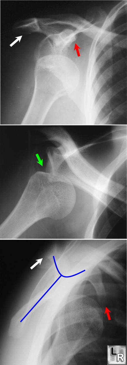
Anterior Dislocation of the Humeral Head: Top image shows humeral head displaced from glenoid
and lying inferior to the coracoid process (red arrow); the middle image demonstrates a defect along the posterolateral aspect of the head, which is the Hill-Sach's deformity (green arrow). The lower image is the scapular Y view (blue line outlines scapula). The head lies in a subcoracoid (i.e. anterior location).
The white arrows point to the acromion.
Emphysematous Cholecystitis

  • General considerations
      • Acute infection of gallbladder caused by gas-forming organism
        • In about 1/3 = clostridium perfringens
        • Also E. Coli and Klebsiella
      • Rare – only 1% of all cases of acute cholecystitis
      • Occurs more often in men
        • As opposed to gallbladder disease in general which occurs more often in women
      • Mostly are elderly patients (>60) with diabetes
      • Vascular compromise of the cystic artery may play a role in the etiology
        • Gallstones may be associated with the disease but are not thought to cause it
      • Gas may occur in the wall and/or the lumen
        • May spread to pericholecystic tissue
        • Rarely, gas may escape into the bile ducts
          • This is rare since cystic duct is usually occluded in cholecystitis
    • Clinical findings
      • As with cholecystitis, right upper quadrant (RUQ) pain and tenderness
      • Leukocytosis
      • Jaundice is rare
    • Imaging findings
      • Conventional radiography
        • May show air in the wall or lumen of the gallbladder
        • Air-fluid levels in the gallbladder will only be seen with images obtained with a horizontal beam, not on supine radiographs
        • Gas may spread to the pericholecystic tissues
        • These findings, if present on the conventional radiograph, usually herald a poor outcome from late-stage disease
      • US findings  
        • Indistinct shadowing emanating from wall or lumen of gallbladder
        • “Ring-down effect” or “comet tail” from shadowing from air in gallbladder lumen
      • CT findings of cholecystitis
        • Air in gallbladder wall is diagnostic of this disease
        • Most common signs of non-emphysematous cholecystitis are gallbladder wall thickening >3mm, and
        • Cholelithiasis
        • Increased density of bile (>20 H)
        • Loss of clear definition of gallbladder wall
        • Pericholecystic fluid such as a halo of edema
    • Treatment
      • Definitive care involves surgical intervention
      • Preoperative percutaneous drainage may improve survival
      • Emergency cholecystectomy
        • Mortality of 15-25%
    • Complications
      • Fivefold increase in perforation over uncomplicated acute cholecystitis
    • Perforation of the gallbladder
      • Frequency is declining because of earlier diagnosis of acute cholecystitis
      • Diagnosis
        • Pre-perforation conventional radiograph showing stones clustered in gallbladder may subsequently show stones scattered in RUQ after perforation
        • Pericholecystic fluid collection on CT or US (not-specific)
        • Scintography may show radiotracer outside of gallbladder in Morrison’s pouch or flank
      • Treatment
        • Preoperative percutaneous drainage of gallbladder and biloma
        • Emergency surgery
  •  

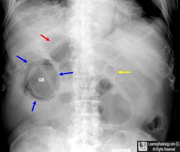
    Emphysematous Cholecystitis

    Emphysematous Cholecystitis. Supine view of the abdomen shows air in the wall (blue arrows) of the gallbladder (GB). There is also a lucency within the lumen of the gallbladder (GB) suggesting air inside the lumen. There is no air-fluid level visible because this radiograph is obtained supine with a vertical x-ray beam. Just superior to the gallbladder is another collection of air (red arrow) that represents a pericholecystic abscess. The yellow arrow points to the end of a PEG tube in the stomach.
    - See more at: http://www.learningradiology.com/archives2014/COW%20590-Emphysematous%20cholecystitis/emphycholecorrect.html#sthash.9oOPXKwD.dpuf
    Garland's triad (also known as the 1-2-3 sign or Pawnbrokers sign) is a lymph node enlargement pattern which has been described in sarcoidosis.
    It comprises of:
    1. right paratracheal nodes
    2. right hilar nodes
    3. left hilar nodes
    Involvement of right paratracheal nodes is not magical but rather reflects the ease with which these nodes are identified on plain radiography. Left paratracheal and aorto-pulmonary nodes are also frequently enlarged, but harder to identify 1

    mercredi 29 janvier 2014

    The apple core sign, also known as a napkin ring sign, is most frequently associated with constriction of the lumen of the colon by a stenosing annular colorectal carcinoma
    The coffee-bean sign or kidney bean sign or bent inner tube sign on an abdominal plain film :
    sigmoid - cecum volvulus ( although some authors have also used the term to refer to closed loop small bowel obstructions )

    Abdominal trauma, Shattered spleen, left renal segmental infarction and left hemothorax

    CT bdomen with IV contrast revealed:-
    - Hypodense zones are seen at upper pole of spleen.
    -Hypodense area is seen at the left renal cortex.
    -Hypodense lienorenal pouch.
    -Laceated lower pole of spleen.
    -Sparing the renal pelvic structures and  vascular bed.
    -all that mentioned above consistant with left side abdominal hematoma with splenic laceration ,,and related to grade III.
    The haemosiderin cap sign refers to an MR imaging feature in some spinal tumourswhere a cap of T2 hypointense haemosiderin is above and / or below the tumour due to previous haemorrhage. 
    It is most often associated with spinal cord ependymomas, being seen in 20-33% of these cases 1. The sign however may also be seen in hemangioblastomas and paragangliomasand therefore it is suggestive of but not pathognomonic for ependymoma