vendredi 7 février 2014


Thrombophlébite cérébrale du sinus latéral G et du sinus longitudinal supérieur avec hématome intraparenchymateux pariétal gauche 


On voit bien le thrombus dans le sinus latéral gauche.


Le sinus longitudinal supérieur n'est pas opacifié.


L'hyperpression veineuse est responsable du saignement intra parenchymateux, qui ne contre-indique pas la décoagulation.

jeudi 6 février 2014


Coffee bean sign - an abdominal plain film sign of sigmoid volvulus. The sigmoid colon has twisted on its mesentery becoming a distended closed loop projecting up and away from the left iliac fossa. The two inner bowel wall edges directly oppose each other simulating the central crevice of a coffee bean. This case also demonstrates
Rigler’s sign of free intraperitoneal gas indicating bowel perforation.   


 - a collection of blood between the inner surface of the skull and the outer layer of the dura. It is typically due to meningeal arterial bleeding and very often associated with a skull fracture. The lentiform shape of extradural haematoma is classically taught as the major method of distinguishing it from the typically crescent shaped subdural haematoma, however there are other helpful distinguishing signs. Unlike subdural blood, extradural blood is not able to cross skull sutures and cannot extend along dural reflections (falx and tentorium).
The above case shows the typical appearance of an extradural haematoma, with the hyperdense lentiform shaped blood being associated with a skull fracture (blue arrow) and the haematoma stopping precisely at the coronal suture (yellow arrow). 


Pneumopericardium


Air appears around heart but does not extend above great vessels

Air in the pericardial sac is limited by the normal pericardial reflections. Pneumo-pericardium is often secondary to an infectious process with associated pericardial fluid and thickening, which will produce an air-fluid level on horizontal beam radiographs. Pneumo-pericardium may produce a similar finding continuous diaphragmatic sign
.
Air within the pericardial sac will rise to a nondependent position on decubitus positioning, unlike mediastinal air, which is not mobile.

jeudi 30 janvier 2014


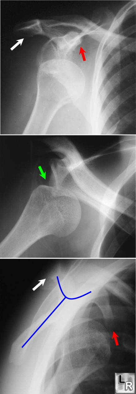
Anterior Dislocation of the Shoulder

·         Glenohumeral dislocation most common shoulder dislocation (85%)
·         Glenohumeral joint dislocations make up >50% of all dislocations in the body
·         Anterior / subcoracoid shoulder dislocation (96%)
·         Mechanism
o       External rotation and abduction and external rotation
·         Age
o       Younger individuals
·         May be associated with
o       Hill-Sachs defect (50%) is a depression fracture of posterolateral surface of humeral head from impaction of the head against glenoid rim in subglenoid type
§         Best demonstrated on the AP projection with the arm internally rotated
o       Bankart lesion is a fracture of anterior aspect of inferior glenoid rim
§         Only cartilaginous portion of glenoid labrum may be fractured which may only be visible on MRI
o       Fracture of greater tuberosity (15%)
·         Complications
o       Recurrent dislocations (40%)
o       Post-traumatic arthritis
o       Injury to axillary nerve or artery


Anterior Dislocation of the Humeral Head: Top image shows humeral head displaced from glenoid
and lying inferior to the coracoid process (red arrow); the middle image demonstrates a defect along the posterolateral aspect of the head, which is the Hill-Sach's deformity (green arrow). The lower image is the scapular Y view (blue line outlines scapula). The head lies in a subcoracoid (i.e. anterior location).
The white arrows point to the acromion.
Emphysematous Cholecystitis

  • General considerations
      • Acute infection of gallbladder caused by gas-forming organism
        • In about 1/3 = clostridium perfringens
        • Also E. Coli and Klebsiella
      • Rare – only 1% of all cases of acute cholecystitis
      • Occurs more often in men
        • As opposed to gallbladder disease in general which occurs more often in women
      • Mostly are elderly patients (>60) with diabetes
      • Vascular compromise of the cystic artery may play a role in the etiology
        • Gallstones may be associated with the disease but are not thought to cause it
      • Gas may occur in the wall and/or the lumen
        • May spread to pericholecystic tissue
        • Rarely, gas may escape into the bile ducts
          • This is rare since cystic duct is usually occluded in cholecystitis
    • Clinical findings
      • As with cholecystitis, right upper quadrant (RUQ) pain and tenderness
      • Leukocytosis
      • Jaundice is rare
    • Imaging findings
      • Conventional radiography
        • May show air in the wall or lumen of the gallbladder
        • Air-fluid levels in the gallbladder will only be seen with images obtained with a horizontal beam, not on supine radiographs
        • Gas may spread to the pericholecystic tissues
        • These findings, if present on the conventional radiograph, usually herald a poor outcome from late-stage disease
      • US findings  
        • Indistinct shadowing emanating from wall or lumen of gallbladder
        • “Ring-down effect” or “comet tail” from shadowing from air in gallbladder lumen
      • CT findings of cholecystitis
        • Air in gallbladder wall is diagnostic of this disease
        • Most common signs of non-emphysematous cholecystitis are gallbladder wall thickening >3mm, and
        • Cholelithiasis
        • Increased density of bile (>20 H)
        • Loss of clear definition of gallbladder wall
        • Pericholecystic fluid such as a halo of edema
    • Treatment
      • Definitive care involves surgical intervention
      • Preoperative percutaneous drainage may improve survival
      • Emergency cholecystectomy
        • Mortality of 15-25%
    • Complications
      • Fivefold increase in perforation over uncomplicated acute cholecystitis
    • Perforation of the gallbladder
      • Frequency is declining because of earlier diagnosis of acute cholecystitis
      • Diagnosis
        • Pre-perforation conventional radiograph showing stones clustered in gallbladder may subsequently show stones scattered in RUQ after perforation
        • Pericholecystic fluid collection on CT or US (not-specific)
        • Scintography may show radiotracer outside of gallbladder in Morrison’s pouch or flank
      • Treatment
        • Preoperative percutaneous drainage of gallbladder and biloma
        • Emergency surgery
  •  

    Emphysematous Cholecystitis

    Emphysematous Cholecystitis. Supine view of the abdomen shows air in the wall (blue arrows) of the gallbladder (GB). There is also a lucency within the lumen of the gallbladder (GB) suggesting air inside the lumen. There is no air-fluid level visible because this radiograph is obtained supine with a vertical x-ray beam. Just superior to the gallbladder is another collection of air (red arrow) that represents a pericholecystic abscess. The yellow arrow points to the end of a PEG tube in the stomach.
    - See more at: http://www.learningradiology.com/archives2014/COW%20590-Emphysematous%20cholecystitis/emphycholecorrect.html#sthash.9oOPXKwD.dpuf
    Garland's triad (also known as the 1-2-3 sign or Pawnbrokers sign) is a lymph node enlargement pattern which has been described in sarcoidosis.
    It comprises of:
    1. right paratracheal nodes
    2. right hilar nodes
    3. left hilar nodes
    Involvement of right paratracheal nodes is not magical but rather reflects the ease with which these nodes are identified on plain radiography. Left paratracheal and aorto-pulmonary nodes are also frequently enlarged, but harder to identify 1